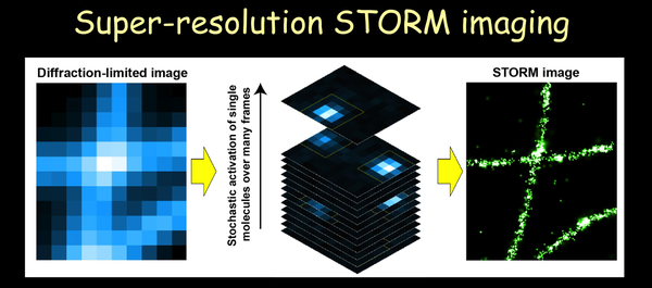
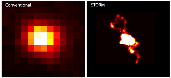
STORM overcomes the diffraction limit by using photo-switchable fluorescent probes to temporally separate the spatially overlapping images of individual molecule. A different subset of fluorescent probes are activated at different times, allowing these fluorophores to be imaged without substantial spatial overlap and to be localized with high precision. Iterating the activation and imaging process allows the position of many fluorescent probes to be determined and a superresolution image to be reconstructed from the positions of the localized probes.
Sub-diffraction-limit imaging by stochastic optical reconstruction microscopy (STORM)
M. J. Rust, M. Bates, X. Zhuang
793-795
(2006)
M. J. Rust, M. Bates, X. Zhuang
Nature Methods
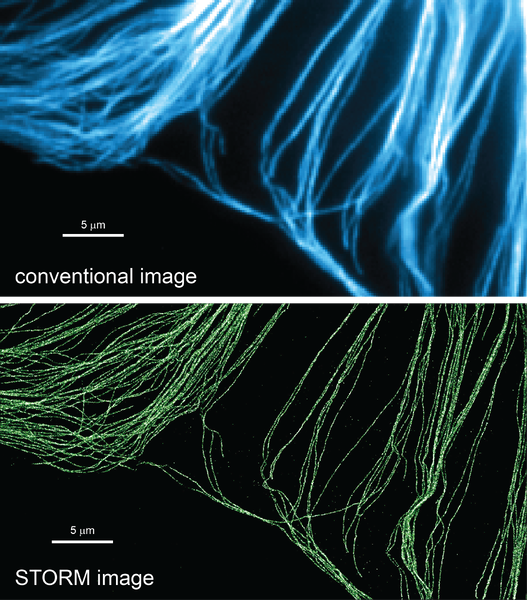
3
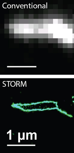
Comparison of conventional diffraction-limited (upper) and STORM (bottom) images of microtubules in a cell. Microtubules are immunolabeled with photoswitchable dyes.
Multicolor Super-Resolution Imaging with Photo-Switchable Fluorescent Probes
M. Bates, B. Huang, G. T. Dempsey, X. Zhuang
1749-1753
(2007)
M. Bates, B. Huang, G. T. Dempsey, X. Zhuang
Science
317
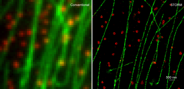
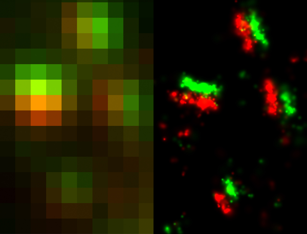
Two-color conventional (Left) and STORM (right) image of microtubules (green) and clathrin-coated pits (red) in a cell. Microtubules and clathrin are immunolabeled with photoswitchable dyes.
Multicolor Super-Resolution Imaging with Photo-Switchable Fluorescent Probes
M. Bates, B. Huang, G. T. Dempsey, X. Zhuang
1749-1753
(2007)
M. Bates, B. Huang, G. T. Dempsey, X. Zhuang
Science
317
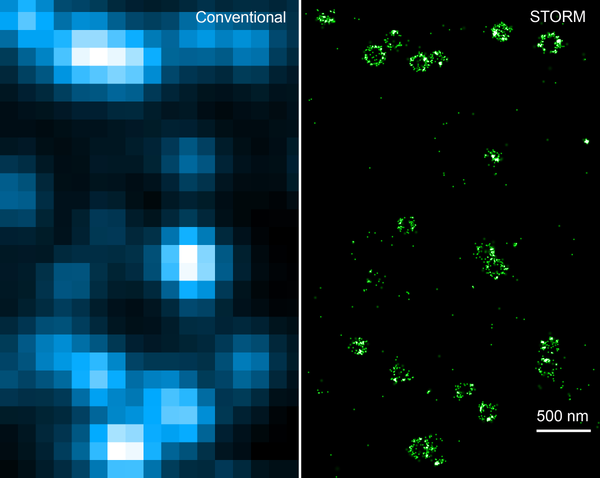
Comparison of conventional (Left) and 3D-STORM (right) images of clathrin-coated pits (red) in a cell. Shown on the right is a xy-cross section of a 3D STORM image. Clathrin is immunolabeled with photoswitchable dyes.
Three-dimensional Super-resolution Imaging by Stochastic Optical Reconstruction Microscopy
B. Huang, W. Wang, M. Bates, X. Zhuang
810-813
(2008)
B. Huang, W. Wang, M. Bates, X. Zhuang
Science
319
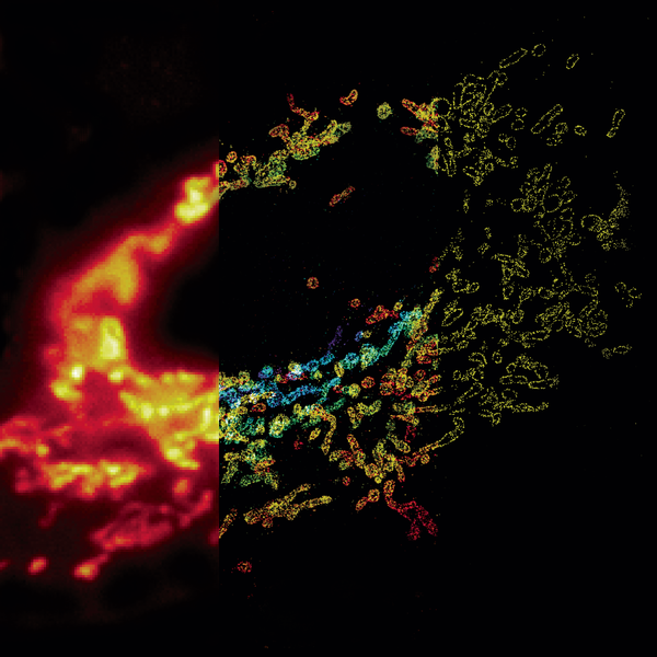
Comparison of conventional and 3D STORM image of mitochondria in a cell. The left panel shows the conventional image of the left 1/3 of the cell. The middle panel shows the 3D STORM image of the middle 1/3 of the cell. The z-dimension information is color-coded. The right panel shows the xy cross-section of the 3D STORM image of the right 1/3 of the cell. TOM20 in on mitochondria is immunolabeled with photoswitchable dyes.
Whole-cell 3D STORM reveals interactions between cellular structures with nanometer-scale resolution
B. Huang, S.A. Jones, B. Brandenburg, X. Zhuang
1047-1052
(2008)
B. Huang, S.A. Jones, B. Brandenburg, X. Zhuang
Nature Methods
5
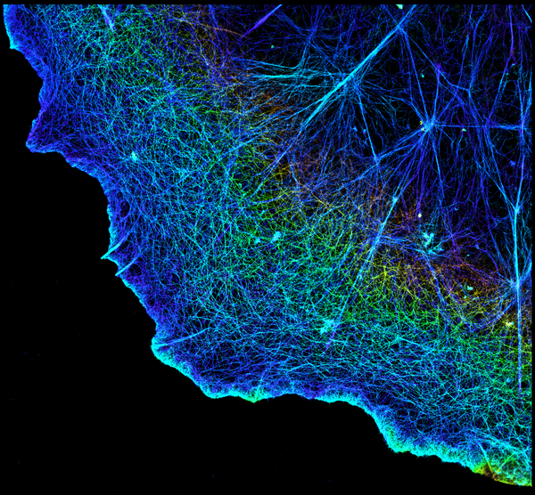
3D STORM image of actin in a cell. The z-dimension information is color-coded. Actin is labeled with phalloidin conjugated to photoswitchable dyes.
Dual-objective STORM Reveals Three-dimensional Filament Organization in the Actin Cytoskeleton
K. Xu, H. Babcock, X. Zhuang
185-188
(2012)
K. Xu, H. Babcock, X. Zhuang
Nature Methods
9
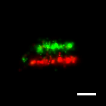
Comparison of conventional (left) and STORM (right) images of chemical synapses in the brain tissue. The synapses are immunolabeled for presynaptic protein Bassoon (red) and postsynaptic protein Homer1 (green) using photoswitchable dyes.
Super-resolution imaging of chemical synapses in the brain
A. Dani, B. Huang, J. Bergan, C. Dulac, X. Zhuang
843-856
(2010)
A. Dani, B. Huang, J. Bergan, C. Dulac, X. Zhuang
Neuron
68
Comparison of conventional (upper) and STORM (bottom) images of telomere loops. The telomere is labeled by fluorescence in situ hybridization with photoswitchable dyes.
Super-resolution fluorescence imaging reveals TRF2-dependent t-loop formation
Y. Doksani, J. Wu, T. de Lange, X. Zhuang
345-356
(2013)
Y. Doksani, J. Wu, T. de Lange, X. Zhuang
Cell
155
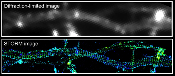
Comparison of conventional (upper) and 3D STORM (lower) images of actin in the axons of neurons. Actin is labeled with phalloidin conjugated to photoswitchable dyes. STORM image revealed a novel periodic, actin-spectrin-based membrane skeleton in axons.
Actin, spectrin and associated proteins form a periodic cytoskeleton structure in axons
K. Xu, G. Zhong, X. Zhuang
452-456
(2013)
K. Xu, G. Zhong, X. Zhuang
Science
339
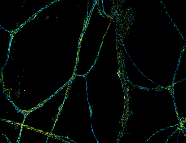
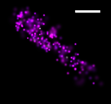
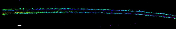
STORM image of a novel periodic, actin-spectrin-based membrane skeleton in axons. This image shows the STORM image of ΒII-spectrin in neuritis immunolabeled with photoswitchable dyes. Except for one neurite in the middle, which is a dendrite, all the other neurities in the image are axons, which exhibit a periodic distribution of ΒII-spectrin.
Actin, spectrin and associated proteins form a periodic cytoskeleton structure in axons
K. Xu, G. Zhong, X. Zhuang
452-456
(2013)
K. Xu, G. Zhong, X. Zhuang
Science
339
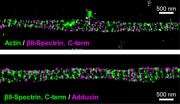
Two-color STORM images of a novel periodic, actin-spectrin based cytoskeleton in axons. The upper panel shows the STORM image of actin (green) and ΒII-spectrin (magenta). The lower panel shows the STORM image of ΒII-spectrin (green) and adducin (magenta). Actin is labeled with with phalloidin conjugated to photoswitchable dyes. ΒII-spectrin is immunostained labeled with photoswitchable dyes against its C-terminal region, which is situated at the center of the spectrin tetramer. Adducin is immunolabeled with photoswitchable dyes.
Actin, spectrin and associated proteins form a periodic cytoskeleton structure in axons
K. Xu, G. Zhong, X. Zhuang
452-456
(2013)
K. Xu, G. Zhong, X. Zhuang
Science
339
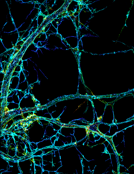
STORM image of a novel periodic, actin-spectrin based cytoskeleton in axons. The image shows the STORM image of actin in neurites. The neurite in the middle is an axon, which shows a periodic distribution of actin. Actin is labeled with phalloidin conjugated to photoswitchable dyes.
Actin, spectrin and associated proteins form a periodic cytoskeleton structure in axons
K. Xu, G. Zhong, X. Zhuang
452-456
(2013)
K. Xu, G. Zhong, X. Zhuang
Science
339
Comparison of conventional (upper) and STORM (bottom) images of chromatin in the nucleus. A specific locus of the chromatin is labeled by fluorescence in situ hybridization with photoswitchable dyes.
Single-molecule super-resolution imaging of chromosomes and in situ haplotype visualization using Oligopaint FISH probes
B. Beliveau, A. Boettiger, M. Avendano, R. Jungmann, R. McCole, E. Joyce, C. Kim-Kiselak, F. Bantignies, C. Fonseka, J. Erceg, M. Hannan, H. Hoang, D. Colognori, J. Lee, W. Shih, P. Yin, X. Zhuang, C.T. Wu
7147
(2015)
B. Beliveau, A. Boettiger, M. Avendano, R. Jungmann, R. McCole, E. Joyce, C. Kim-Kiselak, F. Bantignies, C. Fonseka, J. Erceg, M. Hannan, H. Hoang, D. Colognori, J. Lee, W. Shih, P. Yin, X. Zhuang, C.T. Wu
Nature Communications
6
3D-STORM movie of clathrin-coated pits in a cell. Shown are a series of xy-cross sections of a 3D STORM image. Clathrin is immunolabeled with photoswitchable dyes.
Three-dimensional Super-resolution Imaging by Stochastic Optical Reconstruction Microscopy
B. Huang, W. Wang, M. Bates, X. Zhuang
810-813
(2008)
B. Huang, W. Wang, M. Bates, X. Zhuang
Science
319
3D-STORM movie of ribosomes in a dividing bacterial cell. Shown are a series of xy-cross sections of a 3D STORM image. Ribosome is labeled by fluorescence in situ hybridization with photoswitchable dyes.
Chromosome Organization by a Nucleoid Associated Protein
W. Wang, G. Li, C. Chen, X. Xie, X. Zhuang
1445-1449
(2011)
W. Wang, G. Li, C. Chen, X. Xie, X. Zhuang
Science
333
3D STORM movie of a chemical synapse in the brain tissue. The synapses are immunolabeled for presynaptic protein Bassoon (red) and postsynaptic protein Homer1 (green) using photoswitchable dyes.
Super-resolution imaging of chemical synapses in the brain
A. Dani, B. Huang, J. Bergan, C. Dulac, X. Zhuang
843-856
(2010)
A. Dani, B. Huang, J. Bergan, C. Dulac, X. Zhuang
Neuron
68
3D STORM movie of a calcium channel, CatSper, in the sperm tail. CatSper is immunolabeled with photoswitchable dyes.
Structurally distinct Ca2+ signaling domains of sperm flagella orchestrate tyrosine phosphorylation and motility
J. Chung, S. Shim, R. Everley, S. Gygi, X. Zhuang, D. Clapham
808-822
(2014)
J. Chung, S. Shim, R. Everley, S. Gygi, X. Zhuang, D. Clapham
Cell
157
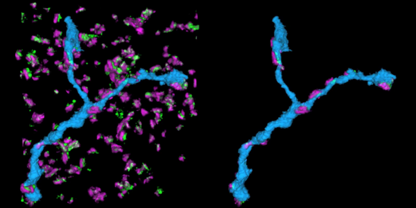
Volumentric STORM movie of dendritic branch immerse in inhibitory synapses. The synapses are immunolabeled for presynaptic proteins (red) and gephrin (green). The neurones express GFP and is immunolabeled using anti-GFP using photoswitchable dyes. The left side of the movie shows an dendritic branch (cyan) amidst synapses, and the right side shows only the synapses assigned to the dendritic branch.
Mapping Synaptic Input Fields of Neurons with Super-Resolution Imaging
Y.M. Sigal, C.M. Speer, H.P. Babcock, X. Zhuang
493-505
(2015)
Y.M. Sigal, C.M. Speer, H.P. Babcock, X. Zhuang
Cell
163
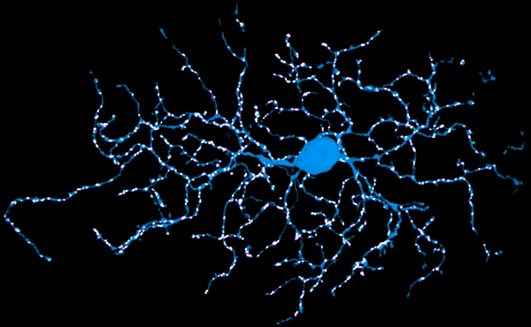
Volumentric STORM movie of a direction-selective retinal ganglion cells with all of its inhibitory input synapses. The synapses are immunolabeled for presynaptic proteins (red) and gephrin (green). The neuron expresses GFP and is immunolabeled using anti-GFP using photoswitchable dyes.
Mapping Synaptic Input Fields of Neurons with Super-Resolution Imaging
Y.M. Sigal, C.M. Speer, H.P. Babcock, X. Zhuang
493-505
(2015)
Y.M. Sigal, C.M. Speer, H.P. Babcock, X. Zhuang
Cell
163